Sample-Scanning
High-Speed Atomic Force Microscope
SS-NEX - Ando model -
Dynamic Visualization of nano-scale world

 |
 |
|
Walking myosin V
N. Kodera et al. 2010 |
Rotorless F1-ATPase
T. Uchihashi et al. 2011 |
Atomic Force Microscope(AFM) is a powerful visualization tool. It's capable imaging real nano-scale structure in air and liquid.
But serious drawback to conventional AFM is slow scanning speed. So that the image is only still picture.
Our High-Speed AFM can observe real-time imaging as movie.
Even swaying samples in solution can be imaged clearly without blurring, since the image acquisition time is short enough.
It is unnecessary to anchor the sample tightly onto substrate, thus the adverse effect from sample preparation is minimized.
*The High-Speed AFM was developed by Prof. Ando (Kanazawa Univ.) and commercialized by RIBM.
 |
 |
|
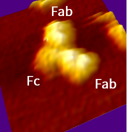
Immunoglobulin G
|
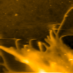
HeLa cell
|
[ Page Top ]
- High scanning rate are archived by unique damping system
- Independent triaxial piezo actuators make images without distorsions
- You can choice one of three type scanners
|
Standard scanner |
Suitable for high- speed imaging such as enzyme reactions and structural changes of protein
|
|---|---|
|
Wide scanner |
Large samples with a high scanning rate
|
|
Mechanically amplified |
Suitable for observing whole body of large samples such as cells
|
-
Note1: The scan range of the scanner is a typical value.
-
Note2: Scan speed is determined for each scanner under specific conditions, and it does not guarantee the scan speed at the maximum scan range.
- Ultra-small cantilever copes with both low spring constant and high resonance frequency
- High-speed scanning with less damage to samples
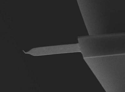
Ultra small cantilever (Olympus)
Resonance Frequency: 1500 kHz (in air), 500 kHz (in liquid)
Spring Constant: 0.1 N / m
Length: 10 μm
- Adopting our unique dynamic PID in addition to wide bandwith analog feedback
- Achieved precize sample imaging without parachuting even in high-speed scanning
[ Page Top ]
| Scan speed | 50 ms / frame (20 frames / sec) |
|---|---|
| Maximum scan range | X: 0.7 μm, Y: 0.7 μm, Z: 0.4 μm |
| Sample size | 1.5 mm in diameter |
| Detection method | Optical lever method |
| Scanning method | Sample scan |
| Environment | In air / In liquid, observation solutions can be added while continuing AFM observation. |
| Control system | PID control, Dynamic PID control |
| Measurement mode | AC mode. Topography, error and phase image |
| Significant function | Scanner active damping, Drift correction for cantilever excitation |
| Software |
|
-
Note1: The scan range of the scanner is a typical value.
-
Note2: Scan speed is determined for each scanner under specific conditions, and it does not guarantee the scan speed at the maximum scan range.
-
Note3: Regarding the options, taking into account on the user needs, the required set up will be arranged by visiting any time.
[ Page Top ]
| Liquid perfusion unit |
Observation solution can be exchanged while continuing AFM observation. |
|---|---|
|
Temperature control unit |
Observation solution can be heated, from room temperature to 40℃. |
|
Illumination system |
Light source: Halogen lamp and mercury lamp(Variable wavelength: 313 - 580 nm) |
[ Page Top ]